The content on this website, including text, graphics, and other materials, is provided for informational purposes only. It is not intended as advice or guidance. Regarding your specific medical condition or treatment, please consult your healthcare provider.
Chronic purulent middle otitis media (CPMO) is an inflammation of the middle ear characterized by persistent perforation (more than 3 months), hearing loss and periodic episodes of otorrhea. Depending on the course and localization of the process, a distinction is made between mesotympanal (mesotympanitis) and atticoantral/epitympanoantral (epitympanitis) purulent otitis media.
Classification
Mesotympanitis, exacerbation;
Mesotympanitis, remission;
Epitympanitis, exacerbation;
Epitympanitis, remission;
Epitympanitis with cholesteatoma of the taut part;
Epitympanitis with cholesteatoma of the unstretched part;
Epitympanitis with polyp.
Mesotympanitis
Etiology of mesotympanitis
A number of factors are necessary for the development of this disease. The primary cause is most often the presence of acute purulent otitis media with a history of perforation of the tympanic membrane.
If auditory tube block persists, inadequate treatment of otitis media, recurrent upper respiratory tract infections, acute otitis media can become chronic.
Less frequently, the cause of CGSA is traumatic or iatrogenic perforation of the tympanic membrane with bacterial infection.
It should be noted that the microbial landscape in patients with CGSA differs from those with acute form of the disease. Most often isolated Pseudomonas aeruginosa и Staphylococcus aureus. These microorganisms are able to form biofilms on the mucosa, through which antibacterial drugs do not penetrate well.
Anatomy of mesotympanitis
This form of chronic purulent otitis media is favorable, since the pathological process is limited to the mucosa and very rarely leads to bone caries. In the course of the disease distinguish periods of exacerbation and remission. The process continues after acute inflammation of the tympanic cavity with perforation of the tympanic membrane. Inflammation affects the mucous membrane of the tympanic cavity in the area of the mesotympanum and hypotympanum, the auditory ossicles and temporal bone are intact. The tympanic membrane is perforated in the stretched part, the edges of the perforation are omosolic, dense, not capable of independent repair, do not affect the fibrous ring. With a prolonged course with frequent recurrences, scarring of the auditory ossicle chain occurs.
The leading complaint of patients is hearing loss on the affected side, up to complete deafness. Periodically , patients note purulent discharge from the external auditory canal, the frequency and duration are highly variable. The discharge is creamy, white-yellow or mucous, there may be an admixture of blood, usually odorless. Painful sensations are extremely rare. In some cases, patients notice a murmur in the ear.
Diagnosis of mesotympanitis
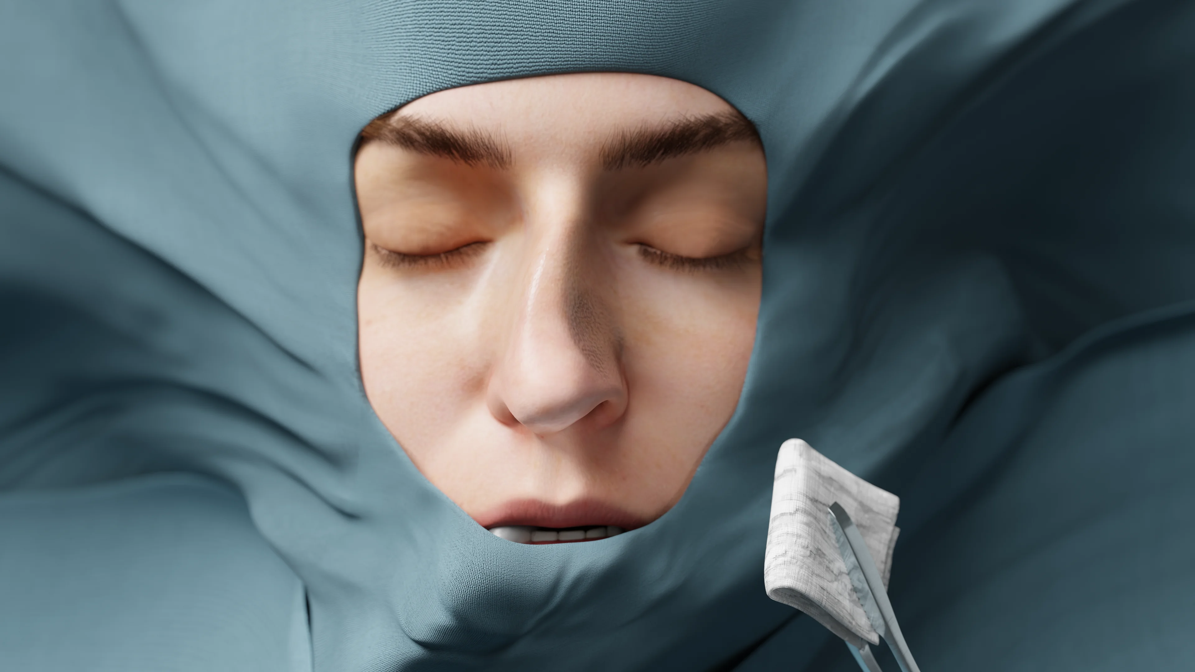
Otomicroscopy is performed first, which reveals a perforation, usually of large size, in the tension portion.
In the remission period, the remnants of the tympanic membrane are gray, the edges of the perforation are clean, a chain of auditory ossicles is visible in the tympanic cavity, and the mucous membrane has a gray tint. The external auditory canal is unchanged.
When the process is exacerbated in the external ear canal, there is abundant purulent content, inflamed and macerated skin of the ear canal, which leads to the development of otitis externa.
The remnants of the tympanic membrane are hyperemic, thickened, full blood, the observable tympanic cavity is also hyperemic.
Mesotympanitis, Exacerbation (Otoscopy) – 3D Model
The degree of patency of the auditory tubes is assessed, and if necessary, a nasopharyngeal endoscopy is performed. A CT scan of the temporal bones is performed to determine the degree of middle ear damage. In order to clarify the degree and nature of hearing loss , chamber tone tests and tonal audiometry are performed.
In case of exacerbation and the presence of pathological discharge, microbiological examination with determination of the causative agent and sensitivity is carried out.
Treatment of mesotympanitis
Conservative and surgical treatment is used for this form of the disease.
Conservative therapy is temporary and does not cure the patient completely. During an exacerbation, the ear canal and tympanic cavity are sanitized, local use of antibacterial drugs. Systemic antibacterial drugs are prescribed with a pronounced course. Concomitant treatment of the upper respiratory tract (to control swelling of the auditory tube) is obligatory.
Surgical intervention is performed for final cure or in case of complications. Scheduled surgical treatment is performed in case of persistent remission (more than 6 months), preserved function of the auditory tube. The main objective of the surgical method is the sanation of the tympanic cavity and reconstruction of the affected tissues. Type 1, 2 or 3 tympanoplasty is performed depending on the volume of the lesion.
If complications develop, emergency extended radical surgery with exposure of the dura or sinuses is performed.
The causes of this type of CGSA are similar to those of mesotympanitis.
Anatomy of epitympanitis
Epitympanitis is a malignant form of CPSO, in which bone tissue is affected along with the mucous membrane. The pathologic process extends to all floors of thetympaniccavity, with predominant lesions of the attic, aditus and antrum. The course of the disease distinguishes between periods of exacerbation and remission. Perforation of the tympanic membrane is localized in its unstretched part, marginal, can be barely distinguishable. The mucous membrane becomes edematous, hyperemic, full of blood. The inflammatory process affects to a greater extent the auditory ossicles, their carious process develops. Subsequently, scar granulations are formed, which cover the affected areas and block the mobility of the chain of auditory ossicles.
Clinical picture of epitympanitis
The insidiousness of this disease lies in the long “mute” period. After some time from the onset of the disease (from several months to several years), scanty stinky purulent discharge (lysed parts of the auditory ossicles) appears. Due to the small size of the perforation in its relaxed part, hearing loss is observed much later, when significant destruction of the ossicles occurs. Patients notice a murmur in the ear, and there may be pain on the affected side during an exacerbation.
Diagnosis of epitympanitis
Otoscopy is obligatory. In case of exacerbation of the process, purulent content with a foul odor is noted in the external auditory canal emanating from a perforation in the relaxed part of the tympanic membrane. The tympanic membrane itself is thickened, turbid pink, the contours are poorly visualized.
During remission, there may be difficulties with diagnosis, especially in the initial stages, as the perforation is located in the non-tensioned part and may be small in size (pinpoint), and the true extent of the lesion behind the tympanic membrane is not visualized.
CT scan of temporal bones is recommended to determine the volume and nature of the lesion, which helps in further planning of surgical intervention.
Among routine methods, chambertonetests and tonal audiography are performed to determine the nature and degree of hearing loss. The degree of patency of the auditory tube must be assessed.
If there is a pathologic discharge, it is sent for microbiological examination to determine the causative agent and sensitivity.
Treatment of epitympanitis
In the period of exacerbation, conservative therapy is prescribed, similar to that for mesotympanitis. Surgical treatment is performed to eliminate the focus of infection and preserve (if possible, improve) hearing. Depending on the extent of the lesion and hearing preservation, open radical antromastoidectomyfollowed by mastoidoplasty, conservative-radical surgery with preservation of viable structures of the tympanic cavity, closed atticotomy or atticoantromastoidectomy and tympanoplasty can be performed afterwards.
Epitympanitis with cholesteatoma of the tensioned, unstretched part
The division of cholesteatoma according to its location is related to the peculiarities of pathologic anatomy and physiology.
Anatomy of epitympanitis with cholesteatoma of the tensioned, unstretched portion
Due to the negative pressure in the tympanic cavity with a blocked auditory tube, the edges of the tympanic membrane perforation are retracted (retraction). Retraction pockets into the tympanic cavity are formed. The upper layer of the tympanic membrane is a multilayered squamous epithelium, which is constantly growing and keratinizing, this process continues in the tympanum. The epithelium comes in contact with the inflamed mucoperiosteal tissue of the tympanic cavity, layering occurs and a cholesteatoma matrix is formed, which is subsequently impregnated with cholesterol. Externally, the cholesteatoma looks like a pearl, yellow-white in color with a pearlescent shimmer. Spreading to all possible parts of the tympanum, the cholesteatoma grows into the bone tissue. The inflamed mucoperiosteum has enzymatic activity (synthesizes collagenase), the cholesteatoma itself has invasive growth, all this leads to bone destruction (caries).
Tensioncholesteatoma is characterized by the localization of a marginal perforation in the tension portion of the tympanic membrane, more often in the posterior-upper quadrant. Cholesteatoma sprouts under the malleus and incus to the posterior portions of the tympanic cavity. It causes block of the attic and impaired ventilation of the superior structures. The process often affects the anvil-stem joint.
Epitympanitis with Cholesteatoma of the Stretched Part of the Tympanic Membrane – 3D Model
Non-tensioned cholesteatoma is formed from the relaxed part of the tympanic membrane, with point perforation. It grows into the upper parts of the tympanum, spreading to the attic, aditus and antrum, in severe cases – to the mastoid process.
In both cases, caries can affect the wall of the facial nerve canal, semicircular canals (more often – horizontal), the wall of the sigmoid sinus, leading to formidable complications.
Clinical picture of epitympanitis with cholesteatoma of the tensioned, unstretched part
Stinky discharge from the external auditory canal with cholesteatomic flakes, conductive hearing loss are characteristic. When the process is severe, there is dull aching pain in the affected ear. When the process spreads to the labyrinth, vestibular symptoms (pronounced vertigo, nystagmus) occur. The most common complications are: labyrinth fistula, facial nerve paresis, sinus thrombosis, meningitis.
Diagnosis of epitympanitis with cholesteatoma of the stretched, unstretched part
The general set of examinations is similar to those listed in other types of CGSA.
Otoscopy during exacerbation reveals purulent discharge in the lumen of the ear canal. Cholesteatomic masses are visualized in the tympanic cavity through the central perforation .
Epitympanitis with Tension Cholesteatoma (Otoscopy) – 3D Model
Characteristic features in patients with cholesteatoma are CT signs of mastoid bone destruction, caries of the auditory ossicles, and increased size of the antrum.
DWI-mode MRI is recommended for visualization of cholesteatoma, where the boundaries of the pathologic mass and healthy tissue, as well as its relation to important anatomical entities can be best traced.
Epitympanitis with a polyp
Anatomy of epitympanitis with polyp
This disease is one of the varieties of epitympanointral otitis media. Due to the constant inflammatory process, necrotic changes in the tympanum mucosa, irritation by purulent discharge, granulation tissue overgrowths occur on the mucosa. Granulations are a substrate for the growth of a polyp, which subsequently prolapses into the perforation of the tympanic membrane and protrudes into the external auditory canal.
The polyp is a soft tissue rounded mass red, shiny, producing a mucous discharge.
If the polyp grows significantly, it completely blocks the perforation. This creates a vicious circle, as inflammation is constantly maintained and a mucopurulent discharge is formed, which accumulates in the tympanic cavity. Subsequently, this content can destroy important structures, causing complications (mastoiditis, labyrinthitis, brain abscess). Often the presence of a polyp in the lumen of the perforation is combined with the spread of cholesteatoma in the tympanic cavity. With the cure of inflammatory processes, polyposis tissue can significantly decrease in size.
Clinical picture of epitympanitis with polyp
In this form of the disease, there is a decrease in hearing,pulsating noise in the affected ear. Patients note a pressing pain in the ear. At exacerbation of the process is characterized by abundant mucous or mucopurulent discharge, sometimes with an admixture of blood.
Find more scientifically accurate content on our social media
Subscribe and don’t miss out the latest resources
Diagnosis of epitympanitis with polyp
Otoscopy reveals a red soft tissue roundish mass prolapsing from the tympanic membrane into the lumen of the ear canal, covered with mucous discharge. On palpation it is elastic, painless, may bleed. In the external auditory canal – mucopurulent discharge. The tympanic membrane is turbid, dense, identifying contours are partially visible.
CT and MRI of the temporal bone are recommended for a more detailed evaluation of the location of the polyp and the spread of the inflammatory process in the middle ear.
Routine examination(tonal audiography, chamber tone tests, vestibular function tests, microbiologic examination of secretions) is also performed.
FAQ
1. What is chronic suppurative otitis media and how dangerous is it?
It is a long-term inflammation of the middle ear with perforation of the eardrum, periodic purulent discharge and hearing loss. The disease is dangerous due to the destruction of ear tissue, deterioration of hearing and the development of serious complications.
2. What are the main symptoms of chronic otitis media?
Symptoms include persistent perforation of the tympanic membrane, purulent discharge from the ear, hearing loss, and noise and sometimes pain during exacerbations.
3. What forms of chronic purulent otitis media exist?
Distinguish mesotympanitis – with inflammation of the middle ear mucosa and a relatively favorable course, and epitympanitis – a more severe form with bone involvement, cholesteatoma and polyps.
4. How long can the disease last?
Otitis media is considered chronic when symptoms are present for more than three months. The disease can last for years with alternating periods of remission and exacerbation.
5. How are diagnostics performed?
The main techniques are otoscopy and otomicroscopy to assess perforation and discharge, CT and MRI to assess the extent of inflammation and bone damage. Microbiologic smears are also taken to determine the causative agent.
6. What are the clinical guidelines for the treatment of chronic suppurative otitis media?
A comprehensive approach is recommended: sanitation of the middle ear and external auditory canal, local and systemic use of antibiotics in exacerbations, as well as surgical treatment (tympanoplasty or radical surgery). It is obligatory to carry out concomitant treatment of the upper respiratory tract, as this is a trigger for the development of chronic otitis media. It is important to monitor the condition to prevent complications.
7. Can otitis media be cured without surgery?
Conservative treatment helps reduce inflammation and symptoms, but does not eliminate the disease completely. Surgical intervention is often necessary to restore the structure of the ear and prevent complications.
8. What complications can occur if left untreated?
It is possible to develop mastoiditis, labyrinthitis, facial nerve paresis, meningitis and other serious conditions that threaten health and life.
List of Sources
1.
VOKA Catalog.
https://catalog.voka.io/
2.
Total Otolaryngology—Head and Neck Surgery, Anthony P. Sclafani, Robin A. Dyleski, Michael J. Pitman, Stimson P. Schantz. Thieme Medical Publishers, Inc., 2015. ISBN 978-1-60406-646-3.
3.
Beerbohm H. Diseases of the ear, throat and nose / Hans Berbom, Oliver Kaschke, Thadeus Navka, Andrew Swift; transl. from English. – 2nd ed. – М. : МЕДпресс-информ, 2016. – 776 с. : ill. ISBN 978-5-00030- 322-1.
4.
Tabassom A, Dahlstrom JJ. Epistaxis. [Updated 2022 Sep 12]. In: StatPearls [Internet]. Treasure Island (FL): StatPearls Publishing; 2025 Jan-.
Available from: https://www.ncbi.nlm.nih.gov/books/NBK435997/
5.
Tunkel DE, Anne S, Payne SC, Ishman SL, Rosenfeld RM, Abramson PJ, Alikhaani JD, Benoit MM, Bercovitz RS, Brown MD, Chernobilsky B, Feldstein DA, Hackell JM, Holbrook EH, Holdsworth SM, Lin KW, Lind MM, Poetker DM, Riley CA, Schneider JS, Seidman MD, Vadlamudi V, Valdez TA, Nnacheta LC, Monjur TM. Clinical Practice Guideline: Nosebleed (Epistaxis). Otolaryngol Head Neck Surg. 2020 Jan;162(1_suppl):S1-S38. doi: 10.1177/0194599819890327. PMID: 31910111.
6.
Mylonas S, Skoulakis C, Nikolaidis V, Hajiioannou J. Epistaxis Treatment Options: Literature Review. Indian J Otolaryngol Head Neck Surg. 2023 Sep;75(3):2235-2244. doi: 10.1007/s12070-023-03824-z. Epub 2023 May 8. PMID: 37636777; PMCID: PMC10447774.
7.
Lüdke T, Müller C, Zahnert T. Chronische mesotympanale Otitis media – Teil 1: Diagnostik & konservative Therapie [Chronic mesotympanic Otitis media – Part 1: Diagnosis and Medical Treatment]. Laryngorhinootologie. 2023 Aug;102(8):619-628. German. doi: 10.1055/a-1961-5714. Epub 2023 Aug 3. PMID: 37536333.