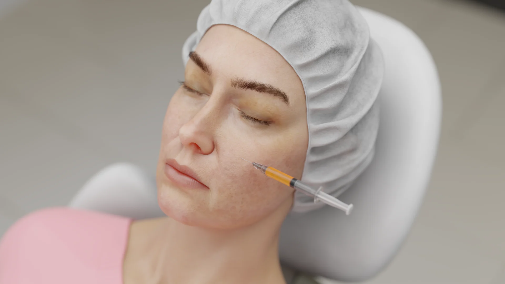
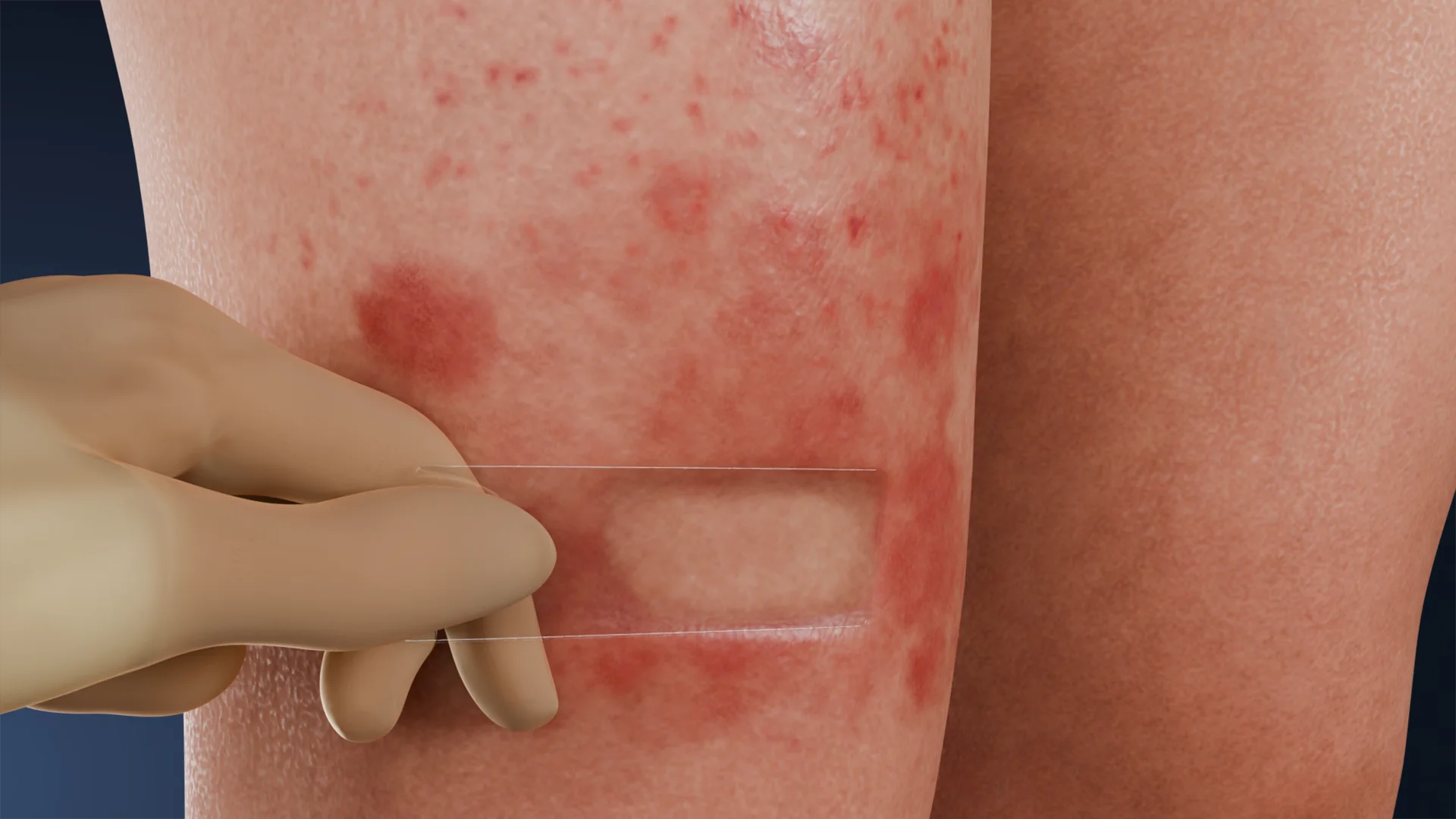
Skin Biopsy in Dermatology: Classification, Technique, and Diagnostic Value
Skin biopsy. This article covers punch, shave, and excisional techniques, specimen handling and fixation, and the role of biopsy in dermatology and oncology.
Anesthesia
Pain management and sedation techniques
Angiology
Arterial and venous pathologies
Cardiology
Acquired and congenital heart diseases
Dentistry
Diseases of teeth, gums, and the oral cavity
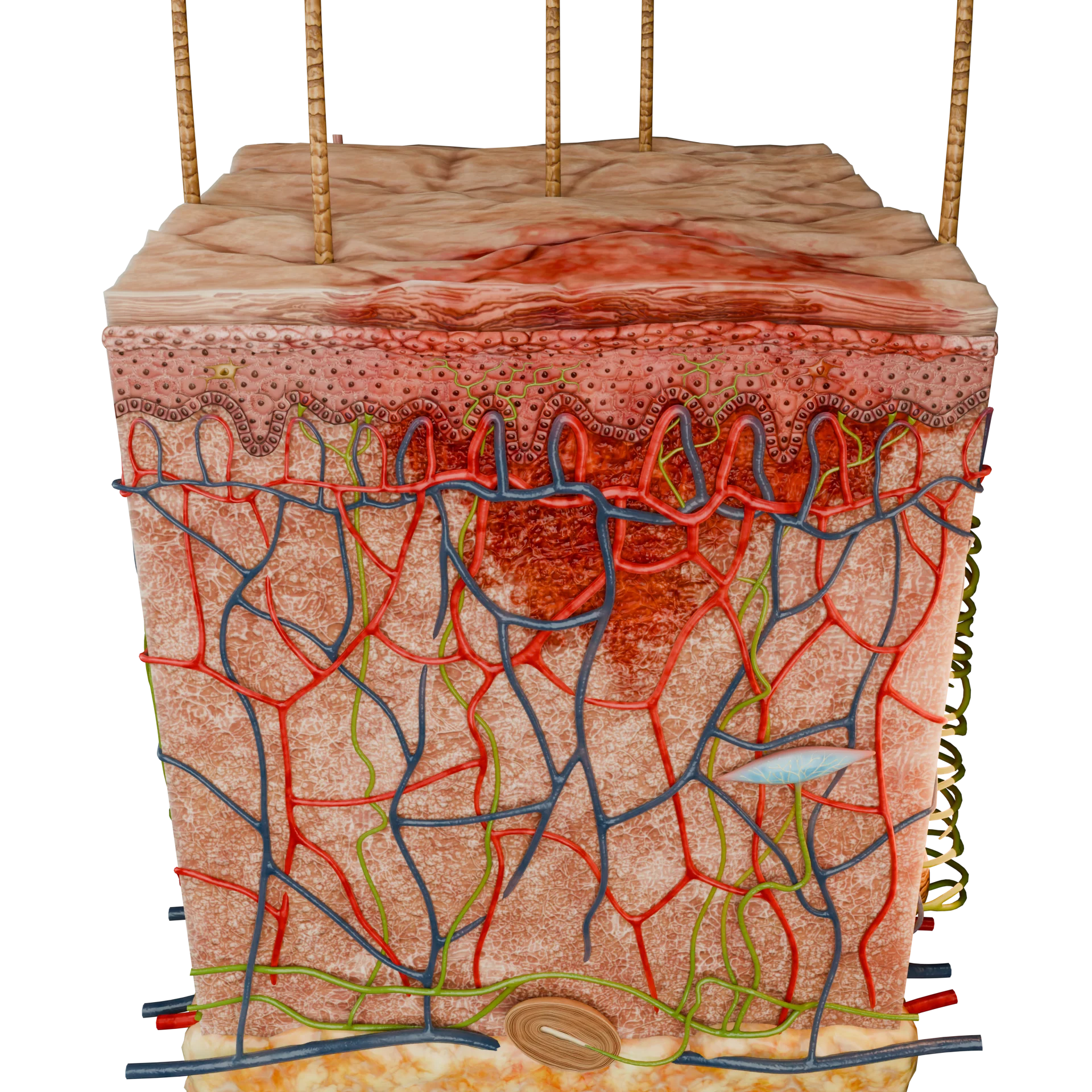
Dermatology
Disorders of the skin and subcutaneous tissue
Endocrinology
Disorders of the glands and hormonal imbalance
Gastroenterology
Stomach, intestinal, and digestive diseases
Gynecology
Diseases of female reproductive organs
Hepatology
Liver, gallbladder, and biliary tract diseases
Neurology
Brain, spinal cord, and peripheral nerve disorders
Obstetrics
Pregnancy complications and abnormal fetal positions
Oncology
Cancer types, benign and malignant tumors
Ophthalmology
Conditions affecting the eyes and vision
Otorhinolaryngology
Ear, nose, and throat diseases
Pediatrics
Child health, development, and clinical conditions
Physiology
Biological processes within organs and systems
Pulmonology
Lung and respiratory tract diseases
Traumatology
Acute injuries and musculoskeletal trauma
Urology
Urinary tract and male reproductive disorders
Anesthesia
Pain management and sedation techniques
Angiology
Arterial and venous pathologies
Cardiology
Acquired and congenital heart diseases
Dentistry
Diseases of teeth, gums, and the oral cavity
Dermatology
Disorders of the skin and subcutaneous tissue
Endocrinology
Disorders of the glands and hormonal imbalance
Gastroenterology
Stomach, intestinal, and digestive diseases
Gynecology
Diseases of female reproductive organs
Hepatology
Liver, gallbladder, and biliary tract diseases
Neurology
Brain, spinal cord, and peripheral nerve disorders
Obstetrics
Pregnancy complications and abnormal fetal positions
Oncology
Cancer types, benign and malignant tumors
Ophthalmology
Conditions affecting the eyes and vision
Otorhinolaryngology
Ear, nose, and throat diseases
Pediatrics
Child health, development, and clinical conditions
Physiology
Biological processes within organs and systems
Pulmonology
Lung and respiratory tract diseases
Traumatology
Acute injuries and musculoskeletal trauma
Urology
Urinary tract and male reproductive disorders

This article is for informational purposes only
The content on this website, including text, graphics, and other materials, is provided for informational purposes only. It is not intended as advice or guidance. Regarding your specific medical condition or treatment, please consult your healthcare provider.
Wood’s lamp is a specialized device used for non-invasive diagnosis of skin diseases, widely applied in cases of skin and hair conditions. This tool was invented in 1903 by physicist Robert Wood, and its first use in dermatology was documented in 1925, when it was recommended for detecting fungal infections.
Originally, the lamp was designed with a special filter that blocked visible light while transmitting only long-wave ultraviolet radiation (UV-A). Modern models employ LED light sources with a wavelength of 365 nm, providing significantly higher power density.

When UV radiation interacts with certain skin conditions, it induces fluorescence — a physical process in which specific substances (endogenous or exogenous) absorb UV-A rays and emit light of a longer wavelength. This reaction is particularly pronounced in various microorganisms. Based on the color of fluorescence, a healthcare professional can make a preliminary assessment of the type, extent, and features of the lesion.
Wood’s lamp is used in dermatology to detect characteristic fluorescence associated with different pathologies.
| Type of lesion | Pathogen / Condition | Characteristic Fluorescence |
|---|---|---|
| Pityriasis versicolor | Pityriasis versicolor (fungus) | Yellow-green |
| Microsporia, dermatophytosis | Fungal infections | Bluish-green / bluish |
| Erythrasma | Corynebacterium minutissimum (bacterium) | Coral-red / red-orange |
| Cutaneous pseudomoniasis | Pseudomonas aeruginosa (bacterium) | Greenish |
| Acne | Cutibacterium acnes (bacterium) | Red-orange (in sebaceous follicles) |
| Vitiligo | Pigmentation disorder | White-blue light |
| Porphyrias | Porphyrin accumulation | Pink-red (blood, urine, enamel) |
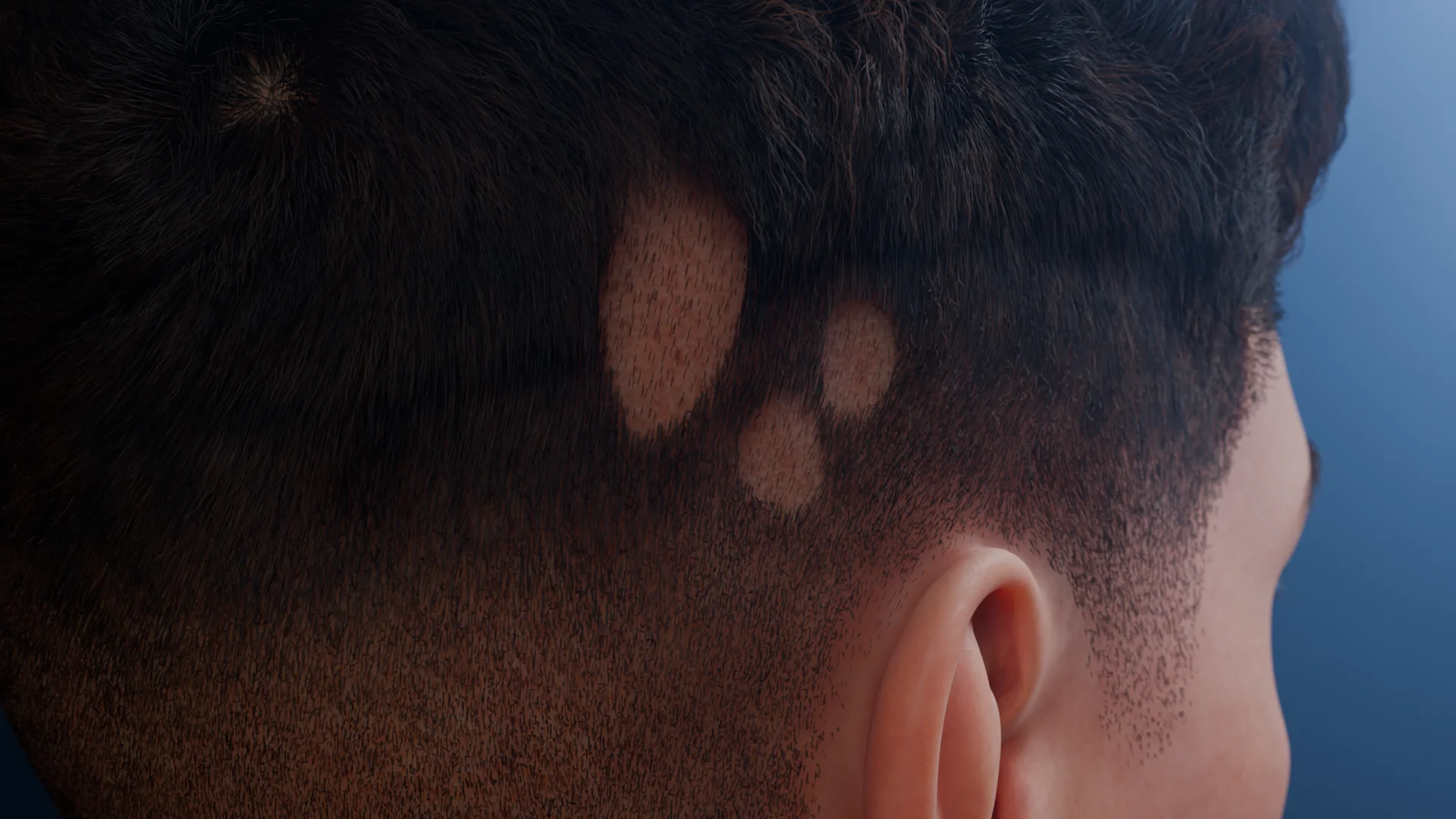 Microsporia Lesion under White Lighting
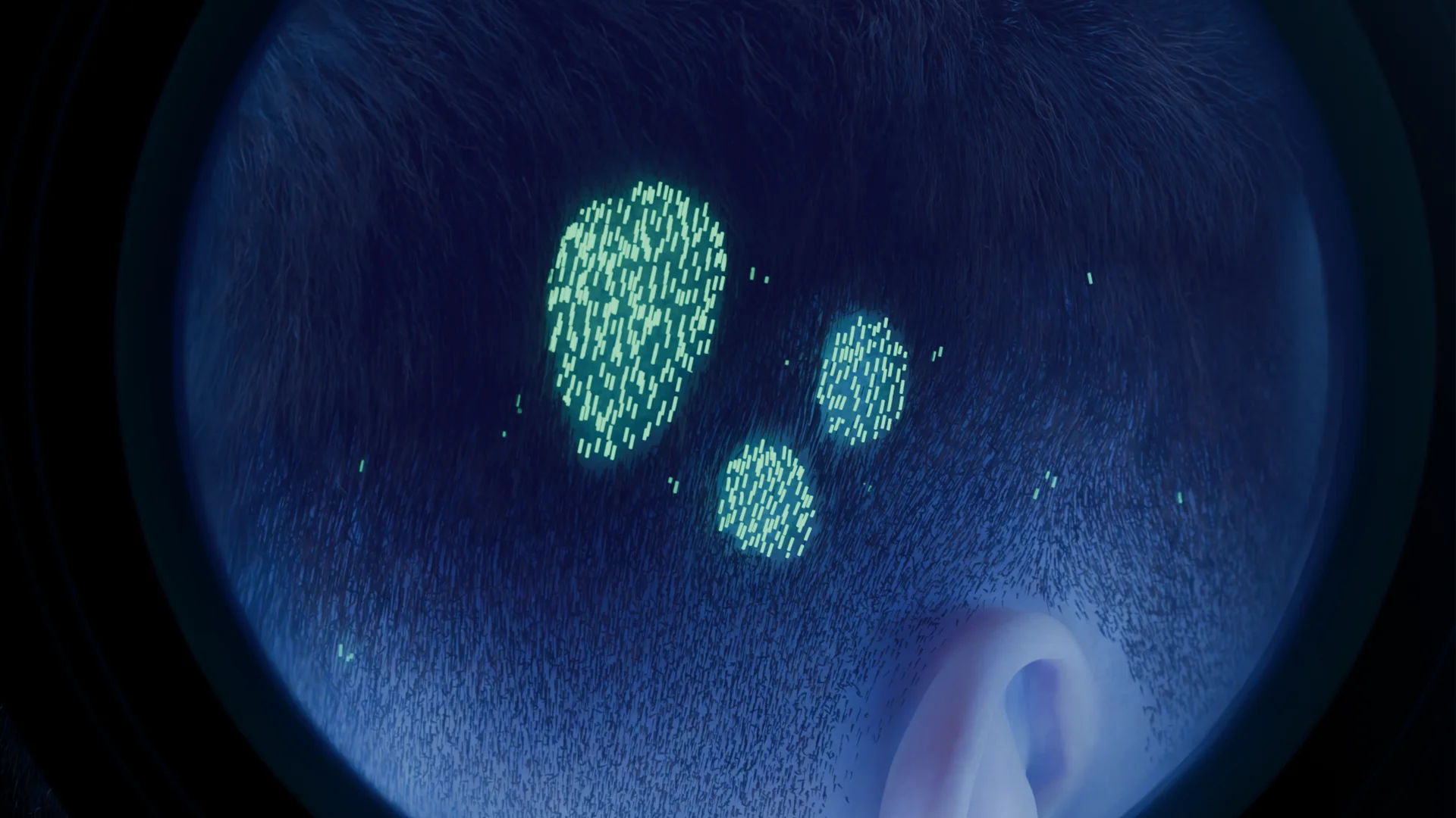 Microsporia Lesion under Wood’s Lamp Illumination
 Pityriasis Versicolor under White Lighting
 Pityriasis Versicolor under Wood’s Lamp Illumination

This is a non-invasive, rapid, and straightforward method.
Find more scientifically accurate content on our social media


Auxiliary role: the method is supplementary and does not replace laboratory diagnostics.
1. How do fungi and pityriasis versicolor fluoresce under Wood’s lamp lighting?
2. What is the purpose of Wood’s lamp in dermatology?
3. What are the key preparation rules prior to Wood’s lamp examination?
4. Can Wood’s lamp harm the skin or eyes?
5. Does Wood’s lamp help determine pigment depth?
6. Why is examination performed in a darkened room?
References
1.
VOKA Catalogue. [Electronic resource].
https://catalog.voka.io/
2.
Kimura Y, Tanemura A, Kurosaki Y, Takafuji M, Yokoi K, Kiyohara E, Arase N, Fujimoto M. Clinical Observation and Proposed Classification of Vitiliginous Patches by a Wood’s Lamp. J Cosmet Dermatol Sci Appl. 2020;10:204–211. doi:10.4236/jcdsa.2020.104021.
3.
Guo W, Qian G, Zhang C. Alopecia from tinea capitis in an 8-year-old boy. CMAJ. 2024 Apr 22;196(15):E526.
4.
Al Aboud DM, Gossman W. Wood’s Light. 2023 Aug 28. In: StatPearls [Internet]. Treasure Island (FL): StatPearls Publishing; 2025 Jan–. PMID: 30725878.
5.
Dyer JM, Foy VM. Revealing The Unseen: A Review of Wood’s Lamp in Dermatology. J Clin Aesthet Dermatol. 2022 Jun;15(6):25‑30
Loading test 6 questions


Summarize article with AI
Choose your preferable AI assistant:
Link successfully copied to clipboard
Thank you!
Your message is sent!
Our experts will contact you shortly. If you have any additional questions, please contact us at info@voka.io