Skin Biopsy in Dermatology: Classification, Technique, and Diagnostic Value
Skin biopsy. This article covers punch, shave, and excisional techniques, specimen handling and fixation, and the role of biopsy in dermatology and oncology.
Anesthesia
Pain management and sedation techniques
Angiology
Arterial and venous pathologies
Cardiology
Acquired and congenital heart diseases
Dentistry
Diseases of teeth, gums, and the oral cavity
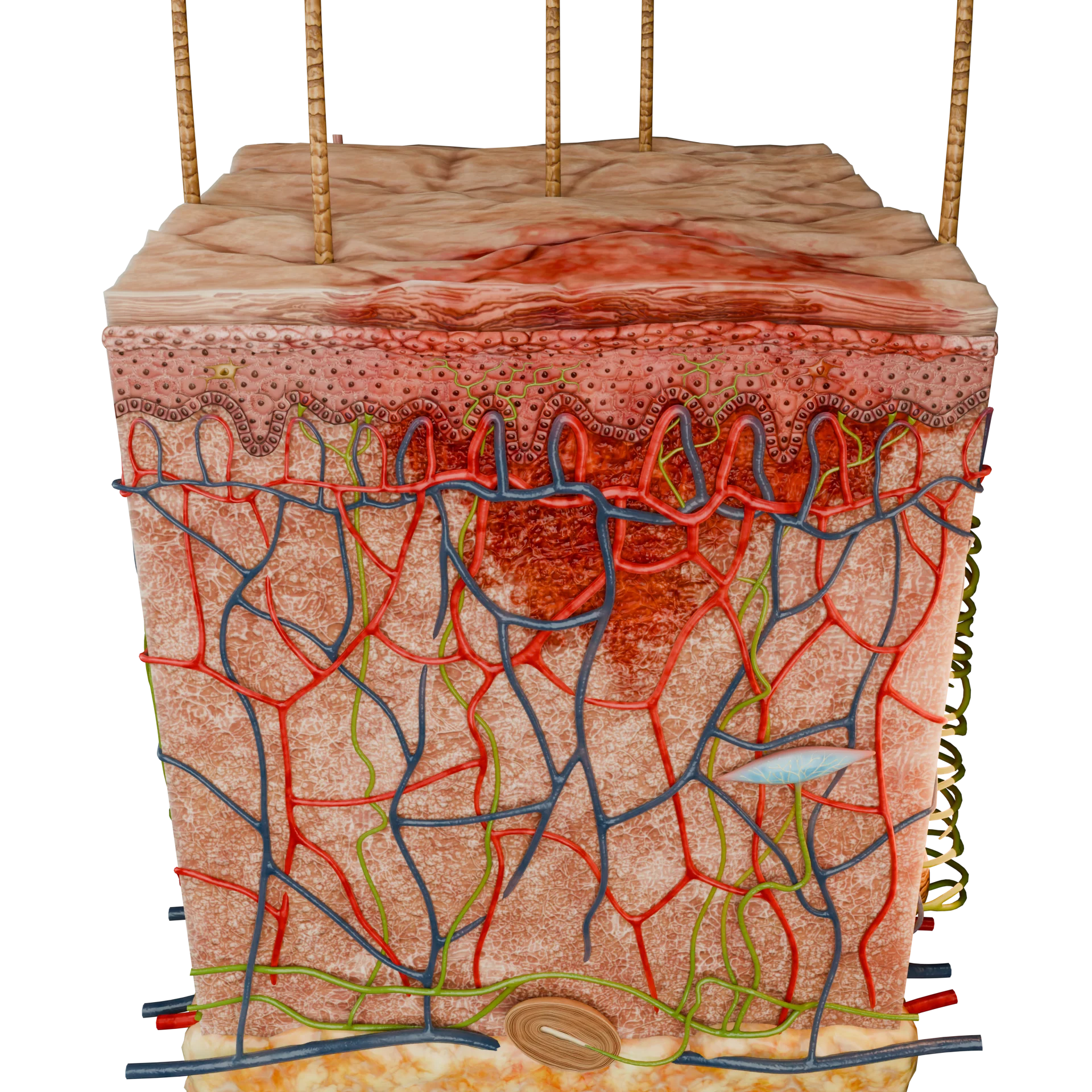
Dermatology
Disorders of the skin and subcutaneous tissue
Endocrinology
Disorders of the glands and hormonal imbalance
Gastroenterology
Stomach, intestinal, and digestive diseases
Gynecology
Diseases of female reproductive organs
Hepatology
Liver, gallbladder, and biliary tract diseases
Neurology
Brain, spinal cord, and peripheral nerve disorders
Obstetrics
Pregnancy complications and abnormal fetal positions
Oncology
Cancer types, benign and malignant tumors
Ophthalmology
Conditions affecting the eyes and vision
Otorhinolaryngology
Ear, nose, and throat diseases
Pediatrics
Child health, development, and clinical conditions
Physiology
Biological processes within organs and systems
Pulmonology
Lung and respiratory tract diseases
Traumatology
Acute injuries and musculoskeletal trauma
Urology
Urinary tract and male reproductive disorders
Anesthesia
Pain management and sedation techniques
Angiology
Arterial and venous pathologies
Cardiology
Acquired and congenital heart diseases
Dentistry
Diseases of teeth, gums, and the oral cavity
Dermatology
Disorders of the skin and subcutaneous tissue
Endocrinology
Disorders of the glands and hormonal imbalance
Gastroenterology
Stomach, intestinal, and digestive diseases
Gynecology
Diseases of female reproductive organs
Hepatology
Liver, gallbladder, and biliary tract diseases
Neurology
Brain, spinal cord, and peripheral nerve disorders
Obstetrics
Pregnancy complications and abnormal fetal positions
Oncology
Cancer types, benign and malignant tumors
Ophthalmology
Conditions affecting the eyes and vision
Otorhinolaryngology
Ear, nose, and throat diseases
Pediatrics
Child health, development, and clinical conditions
Physiology
Biological processes within organs and systems
Pulmonology
Lung and respiratory tract diseases
Traumatology
Acute injuries and musculoskeletal trauma
Urology
Urinary tract and male reproductive disorders

This article is for informational purposes only
The content on this website, including text, graphics, and other materials, is provided for informational purposes only. It is not intended as advice or guidance. Regarding your specific medical condition or treatment, please consult your healthcare provider.
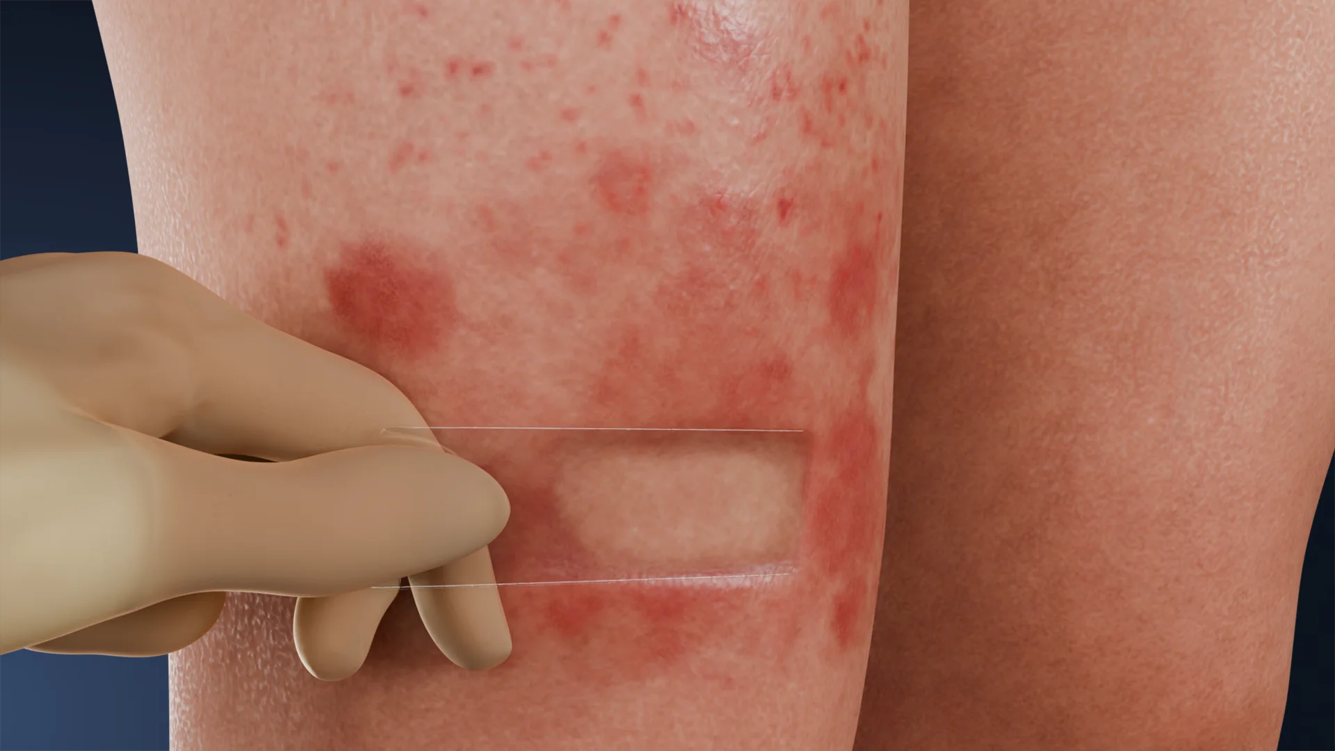
Diascopy is a simple yet informative diagnostic method in dermatology, based on the examination of skin changes under pressure. This technique is still used in modern dermatology due to its high diagnostic effectiveness and accessibility. Diascopy was first described in the late 19th century and remains in clinical use today.
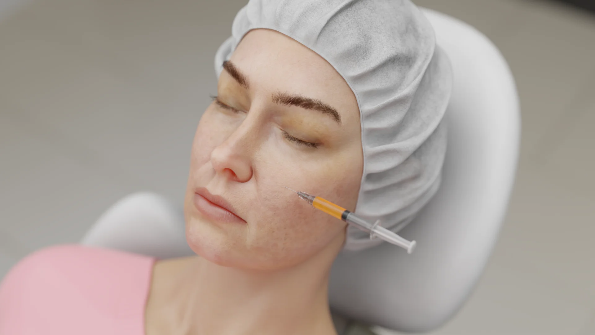
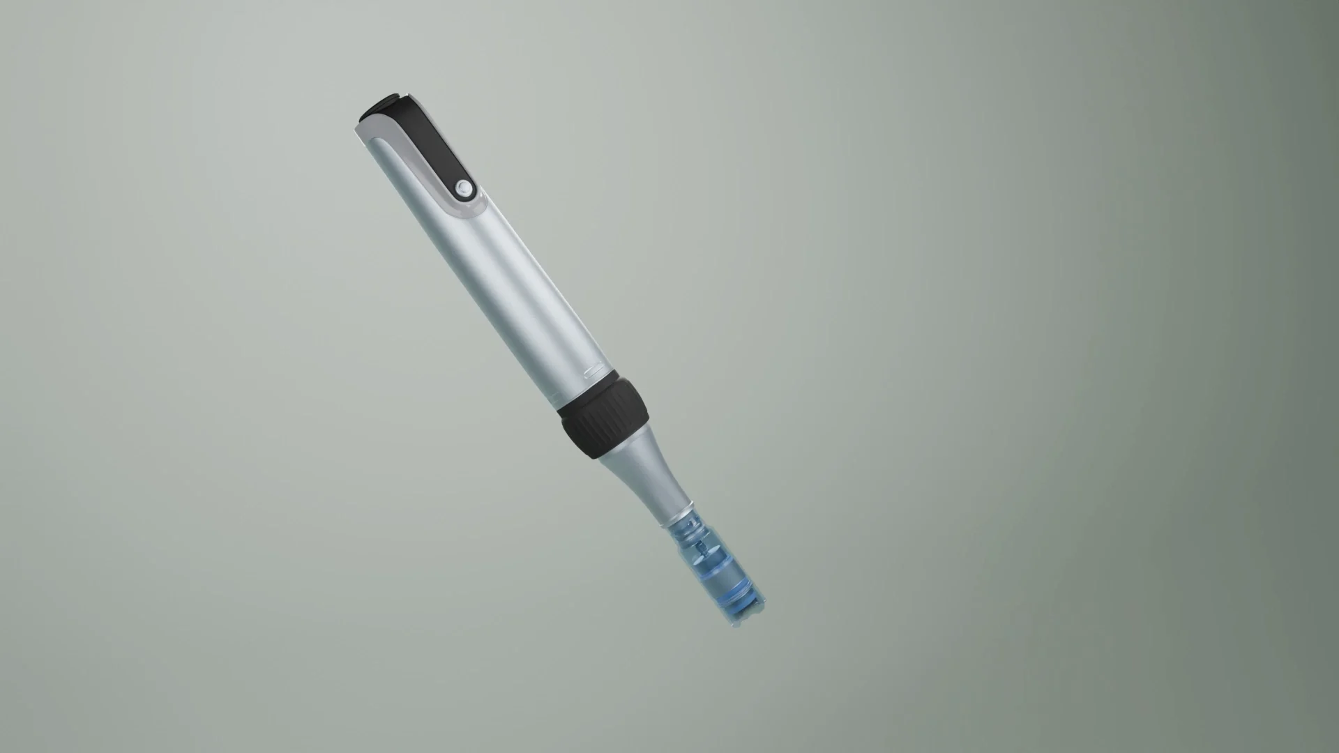
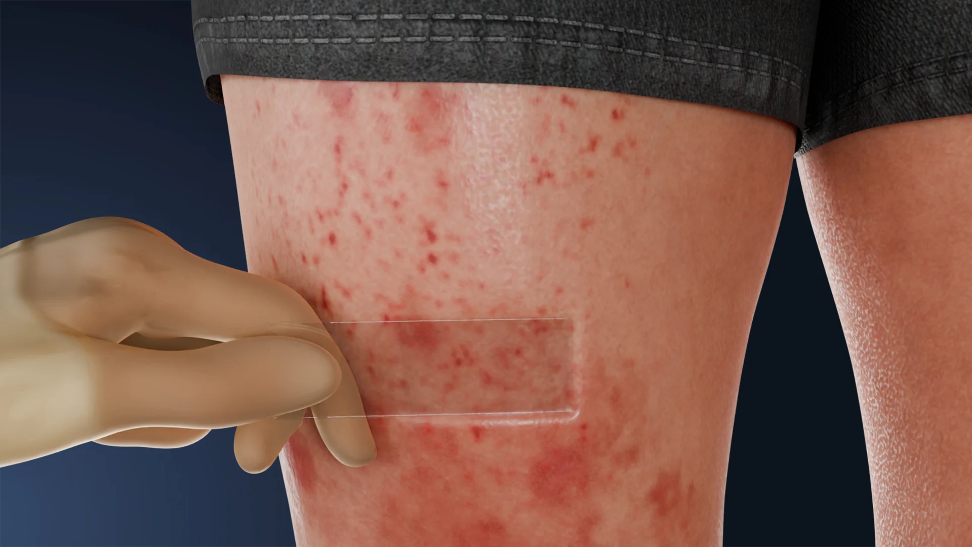
Diascopy is performed by applying light pressure to the skin surface with a transparent glass or plastic spatula. For this purpose, a diascopic device with illumination or a dermatoscope may also be utilized. The procedure is based on the phenomenon of optical clearing of the epidermis under compression, which allows the healthcare professional to visually determine the nature of morphological elements, specifically:
Diascopy is performed in several steps:
The changes observed under pressure are differentiated into:

Find more scientifically accurate content on our social media


1. What are the main indications for diascopy?
2. Can diascopy be used instead of skin biopsy?
3. When is diascopy least informative?
4. What is positive diascopy?
5. When does the “apple jelly” sign apply?
References
1.
VOKA Catalogue. [Electronic resource].
https://catalog.voka.io/
2.
Dermoscopy in general dermatology: practical tips for the clinician British Journal of Dermatology, Volume 170, Issue 3, 1 March 2014, Pages 514–526.
3.
Matos D, Coelho R. “Apple Jelly” Sign: Diascopy in Cutaneous Sarcoidosis. Acta Med Port. 2015 May-Jun;28(3):394. doi: 10.20344/amp.5396. Epub 2015 Jun 30. PMID: 26421796.
4.
Скрипкин, Ю. К. Дерматовенерология. Национальное руководство / под ред. Ю. К. Скрипкина, Ю. С. Бутова, О. Л. Иванова. – Москва : ГЭОТАР-Медиа, 2014. [Skripkin, Yu. K. (2014). Dermatovenerologia. National’noye rukovodstvo (Dermatovenereology: National guidelines) Skripkin, Yu.K. (Ed.), Butov, Yu. S. (Ed.), Ivanova, O. L. (Ed.). Moscow: GEOTAR-Media]. – 1024 с.
5.
Clinical Dermatology: A Color Guide to Diagnosis and Therapy 6th Edition by Thomas P. Habif MD (Author).
6.
Dermatology, 5th Edition. Author : By Jean L. Bolognia, MD, Julie V. Schaffer, MD and Lorenzo Cerroni, MD.
Loading test 6 questions
Table of Contents


Summarize article with AI
Choose your preferable AI assistant:
Link successfully copied to clipboard
Thank you!
Your message is sent!
Our experts will contact you shortly. If you have any additional questions, please contact us at info@voka.io